Post-transplant Cytomegalovirus Infection

Cytomegalovirus (CMV) is one of the most common complications among recipients of solid organ transplants (SOT) and hematopoietic stem cell transplants (HSCT).1,2

Among transplant recipients, CMV infection may lead to CMV end-organ disease, increased morbidity, and higher rates of mortality.1,2

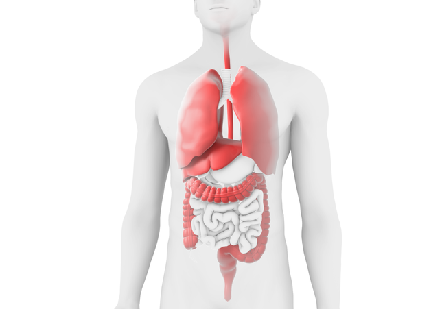
Cytomegalovirus Infection illustration

Epidemiology

The seroprevalence of CMV is high in the general population of the United States, but has a greater risk of clinical consequences when the immune system is suppressed, such as during SOT or HSCT.1,3,4

Nearly 40,000 solid organ transplants were performed in the US in 2020.5 Earlier studies estimate that the incidence of CMV disease ranges from 8-32% in heart, kidney, and liver recipients, and is as high as 75% in lung recipients.6 Risk factors for CMV disease in SOT include serological mismatch, intense immunosuppression, and lung transplant.1,6 D+/R- transplants are associated with the highest risk of CMV disease (19.2% to 31.3%) because seronegative recipients lack cellular and humoral immunity to CMV.7,8,9

There were about 22,000 HSCTs performed in the US in 2020.10 Estimates for the incidence of CMV disease range from 5% in autologous transplants to 30% in allogeneic transplant.6 Risk factors for CMV infection in HSCT include serological mismatch, treatment for GvHD, and treatment with anti-thymocyte globulin. Seropositive HSCT recipients have the highest risk of CMV reactivation. D-/R+ transplants are at highest risk for infection (36%) because the lack of CMV-specific memory T cells prolongs immunological anti-CMV reconstitution.6,11

Diagnosis

CMV infection in transplant recipients is diagnosed through identification of viral replication, which is monitored post-transplant.6,12,13 The most common method is quantitative nucleic acid testing (QNAT), which is a real-time polymerase chain reaction (RT-PCR) method. QNAT, which can be advantageous for standardization, provides quantification of CMV DNA.

Pathophysiology

CMV is a herpesvirus with double-stranded DNA.2 CMV can lie dormant in cells and reactivate when the host is immunocompromised.6,14,15

The seroprevalence of CMV is high in the general population, with transmission via saliva, sexual contact, placental transfer, breastfeeding, transfusion, and transplantation.15

Primary infection involves the lytic life cycle (lysis of the host cell), but is commonly asymptomatic in immunocompetent hosts.14,15 CMV then enters a latent phase in which the virus lies dormant within a host cell, avoiding immune surveillance. Latency is normally asymptomatic. Reactivation is when the virus re-enters the lytic life cycle. This can be triggered by immunosuppression, inflammation, infection, and stress, and commonly occurs in immunocompromised hosts, such as transplant recipients.15

Navigating CMV

In the post-transplant setting, the transplant team must deploy a risk-based testing strategy to guide the approach to CMV prophylaxis, modulation of immunosuppressive regimen, and if necessary, treatment of CMV.6

Adding to the challenge of CMV management is the presence of refractory CMV, defined as a suboptimal response to antiviral therapy; and resistant CMV, a laboratory definition of a drug-resistant phenotype or the presence of mutations known to confer resistance to antiviral agents.16 CMV that is refractory and/or resistant to treatment can lead to high morbidity and mortality rates.16,17 Ongoing and future research looks to improve sequencing and detection of mutations, and identification of optimal therapeutic strategies.16

  1. Kotton CN, Kumar D, Caliendo AM, et al. Transplantation. 2018;102(6):900-31.
  2. El Chaer F, Shah DP, Chemaly RF. Blood. 2016;128(23):2624-2636.
  3. US Department of Health and Human Services, Food and Drug Administration, Center for Drug Evaluation and Research. Cytomegalovirus in Transplantation: Developing Drugs to Treat or Prevent Disease. Guidance for Industry. May 2020. https://www.fda.gov/media/112942/download. Accessed October 24, 2022.
  4. Cho S-Y, Lee D-G, Kim H-J. Int J Mol Sci. 2019;20(11):2666.
  5. HRSA. Organ Procurement and Transplantation Network. Available from: https://optn.transplant.hrsa.gov/news/organ-transplants-in-united-states-set-sixth-consecutive-record-in-2018/. Accessed September 2022.
  6. Azevedo LS, Pierrotti LC, Abdala E, et al. Clinics (Sao Paulo). 2015;70(7):515-523.
  7. Humar A, Snydman D, AST Infectious Diseases Community of Practice. Am J Transplant. 2009;9(Suppl 4):S78-S86.
  8. Harvala H, Steward C, Muller K, et al. J Med Virol. 2013;85(5):893-898.
  9. Mendez-Eirin E, Paniagua-Martin MJ, Marzoa-Rivas R, et al. Transplant Proc. 2012;44(9):2660-2662.
  10. HRSA. Blood Stem Cell. Donation and Transplantation Statistics. Available from: https://bloodstemcell.hrsa.gov/data/donation-and-transplantation-statistics/transplant-activity-report. Accessed September 2022.
  11. Styczynski J. Infect Dis Ther. 2018;7(1):1-16.
  12. Razonable RR, Humar A, AST Infectious Diseases Community of Practice. Am J Transplant. 2013;13
    (Suppl 4):93-106.
  13. Ljungman P, Hakki M, Boeckh M. Hematol Oncol Clin North Am. 2011;25(1):151-169.
  14. Fishman JA. Am J Transplant. 2013;13(Suppl 3):1-8.
  15. Crough T and Khanna R. Clin Microbiol Rev. 2009;22(1):76-98.
  16. Chemaly RF, Chou S, Einsele H, et al. Clin Infect Dis. 2019;68(8):1420-1426.
  17. Fisher CE, Knudsen JL, Jerome KR, et al. Clin Infect Dis. 2017;65(1):57-63.