Gaucher Disease (GD) | Takeda U.S. Medical

Gaucher Disease                     

Gaucher disease is one of the most common lysosomal storage disorders (LSD), in which lipid-filled cells displace normal cells in multiple organs.1,2,3,4

LSDs are a group of more than 70 inherited metabolic diseases marked by lysosomal accumulation of waste products.5 Gaucher Disease (GD), named after French physician Phillipe Gaucher who first described it in 1882, is characterized by the accumulation of glycolipid glucocerebroside (glucosylceramide) in macrophages.6 These lipid-filled Gaucher cells displace normal cells in bone marrow, spleen, liver, lungs and in neuronopathic types in the nervous system as well, resulting in its variable clinical presentation.2,3,4,7

Gaucher disease illustration

Epidemiology

GD is a rare disease with a frequency of about 1 in 50,000 to 1 in 100,000 worldwide, but it’s particularly more common in the Ashkenazi Jewish population (1 in 800 to 1 in 1,000).6,8 Due to its inheritance pattern, it tends to affect males and females equally.3 The onset of symptoms can vary depending on disease type and severity with milder forms presenting in adulthood and a significant number of individuals with type 1 never receiving any medical attention.9,10

GD is generally categorized into 3 types: type 1 is the most prevalent, accounting for more than 90% of all cases, and is characterized by the presence of bone disease and the lack of neurologic features. Type 2 and Type 3 both typically involve neurodegeneration.7,10,11,12,13

Pathophysiology

GD is an autosomal recessive inherited disorder that causes a deficiency in the β-glucocerebrosidase enzyme, which is required for glycosphingolipid metabolism.1,2,3 GD has been associated with >350 different variants in the Glucosylceramidase Beta 1 (GBA1) gene and results in lysosomal accumulation of glucosylceramide within cells, leading to its multisystemic manifestations.6

The manifestations of Type 1 GD are caused by the accumulation of engorged macrophages in visceral tissues.11 By contrast, Types 2 and 3 GD have a pathophysiology that leads to neuronal death, possibly due to the inability to break down glucosylceramide, and the accumulation of a derivative neurotoxin, glucosylsphingosine.6

Diagnosis

Identifying patients for diagnostic testing remains a challenge in clinical practice, contributing to diagnostic delays and misdiagnoses.14,15

Although patients are commonly diagnosed in childhood or adolescence, the average age at diagnosis for the most common type of GD (type 1) is 30-40 years of age.9

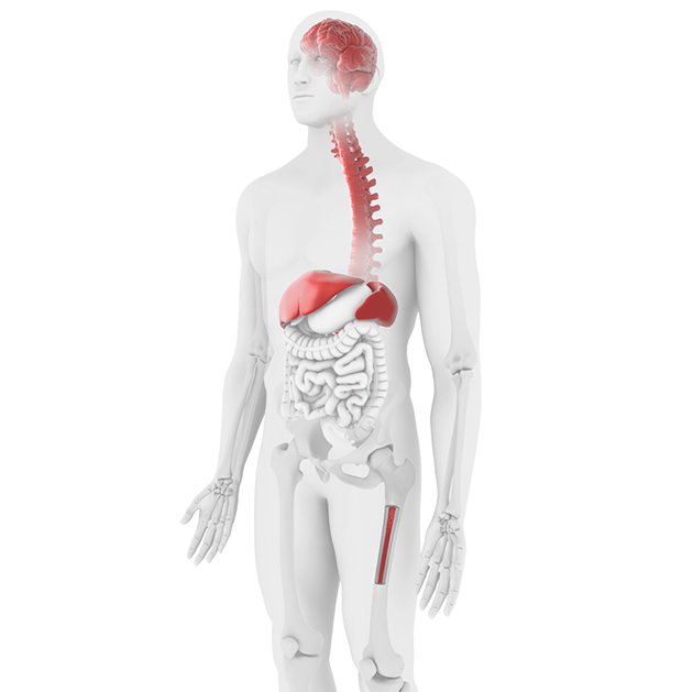
Initial diagnosis is based on laboratory testing and physical examination including:16

  • hematologic (complete blood count, liver function test)
  • skeletal (bone mineral density, magnetic resonance imaging (MRI) of hip/spine)
  • visceral (MRI of spleen/liver)
  • cardiopulmonary (electrocardiogram, chest x-ray)
  • central nervous system (neurological exams)

However, a definitive diagnosis requires enzymatic analysis of glucocerebrosidase and can also be supplemented by detection of genetic defects.17

Type 2 GD, with substantial CNS involvement, is typically diagnosed in infancy, with bulbar signs and oculomotor paresis, while Type 3 GD is typically diagnosed in childhood or adolescence, often with a progression of CNS involvement.11

Navigating Gaucher Disease

Several factors are consistently elevated in GD and can therefore be used as potential biomarkers to:8

  • Aid in the diagnosis and monitoring of disease progression8,18
  • Provide insight into disease pathophysiology19
  • Aid in the development of GD-specific treatments19,20

Traditional biomarkers (chitotriosidase and CCL18) have demonstrated low sensitivity and lack specificity.21 Glucosylsphingosine (Lyso-Gb1) has been associated with 100% sensitivity/specificity, is correlated with liver volume and bone marrow fat fraction, and with severity of variants in the GBA gene.4,18,21

  1. Burrow TA, Barnes S, Grabowski GA. Ped Health Med Therapeutics. 2011;2:59-73.
  2. Fuller M. Lipids Health Dis. 2010;9:113.
  3. Grabowski GA, Petsko GA, Kolodny EH. Gaucher Disease. In: Valle D, et al. eds. The Online Metabolic and Molecular Bases of Inherited Disease. New York, NY: McGraw-Hill; 2014. https://ommbid.mhmedical.com/content.aspx?bookid=2709&sectionid=225546056. Accessed October 28, 2022.
  4. Dekker N, van Dussen L, Hollak CE, et al. Blood. 2011;118(16):e118-e127.
  5. Platt FM, d'Azzo A, Davidson BL, et al. Nat Rev Dis Primers. 2018;4(1):27.
  6. Grabowski GA. Hematology. 2012;2012:13-18.
  7. Weinreb NJ and vom Dahl S. Blood. 2008;112(11):3549.
  8. Mistry PK, Cappellini MD, Lukina E, et al. Am J Hematol. 2011;86(1):110-115.
  9. Ferreira CR and Gahl WA. Transl Sci Rare Dis. 2017;2(1-2):1-71.
  10. Mankin HJ, Rosenthal DI, Xavier R. J Bone Joint Surg Am. 2001;83(5):748-762.
  11. Grabowski GA. Lancet. 2008;372(9645):1263-1271.
  12. Koprivica V, Stone DL, Park JK, et al. Am J Hum Genet. 2000;66(6):1777-1786.
  13. Pastores GM, Weinreb, NJ, Aerts H, et al. Semin Hematol. 2004;41(Suppl 5):4-14.
  14. Mehta A, Kuter DJ, Salek SS, et al. Intern Med J. 2019;49(5):578-591.
  15. Mehta A, Belmatoug N, Bembi B, et al. Mol Genet Metab. 2017;122(3):122-129.
  16. Beutler E and Grabowski GA. The metabolic & molecular bases of inherited disease. 8th edn. New York: McGraw-Hill, 2001;3:3635-68.
  17. Salehpour S. Iran J Child Neurol. 2015;9:14-15.
  18. Rolfs A, Oprea G, Kramp G, et al. Mol Genet Metab. 2017;120:S116.
  19. Aerts JM, Kallemeijn WW, Wegdam W, et al. J Inherit Metab Dis. 2011;34(3):605-619.
  20. European Medicines Agency. Report on the EMEA/CHMP Biomarkers Workshop. Available from: www.ema.europa.eu/docs/en_GB/document_library/Report/2009/11/WC500012540.pdf. Accessed August 15, 2022.
  21. Rolfs A, Anne-Katrin G, Grittner U, et al. PLOS One. 2013;8(11):e79732.